FluoroSim

Practise the needle before the patient.

A fluoroscopy simulator for spinal and joint pain procedures. Swing the C-arm, steer the needle, inject contrast — then see how close you came, what you passed on the way, and what the ideal path would have been.

Free. Runs in your browser on a computer or iPad, with nothing to install and no account.
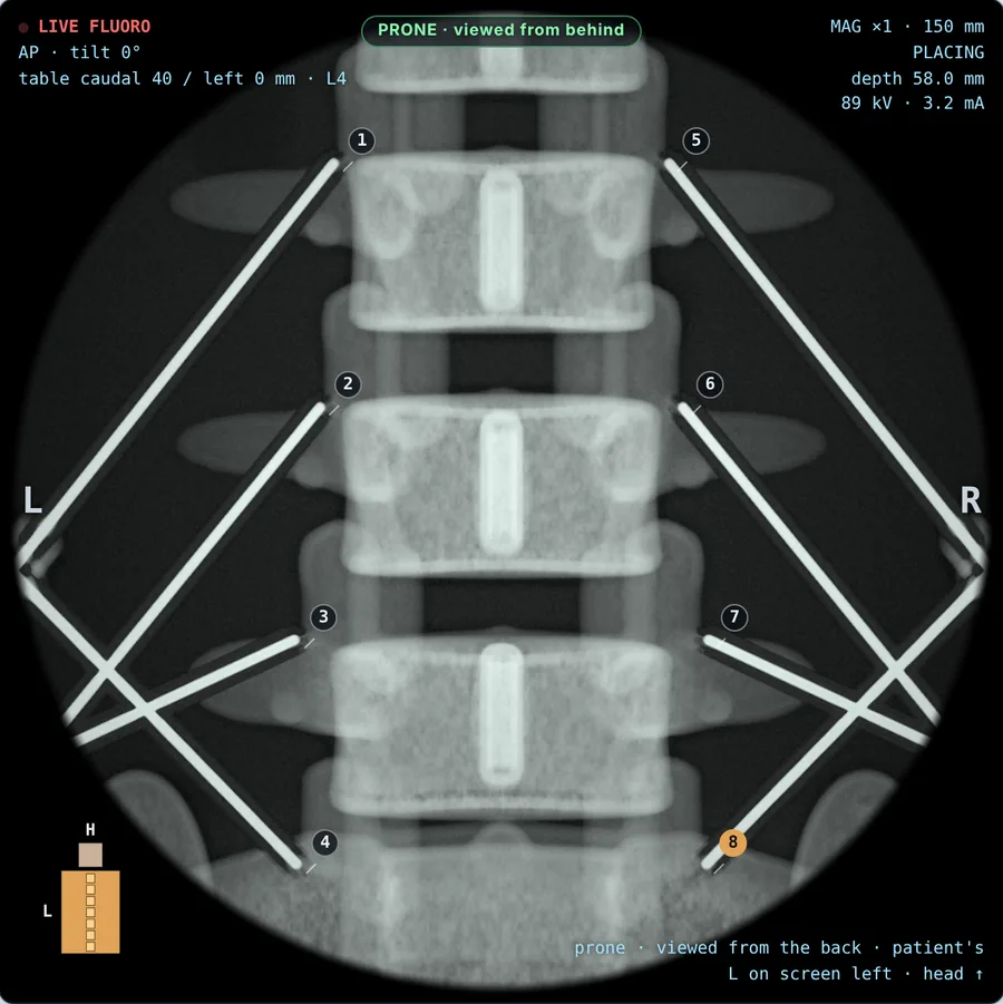
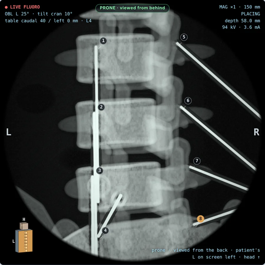
Simulated AP fluoroscopy of the lumbar spine with eight radiofrequency cannulae placed bilaterally at L2 to L5, each tagged with its number.
Last image holdBilateral L2–L5 medial branch RF: eight cannulae, AP view

A whole procedure list, on simulated patients

Every case is a patient you can screen from any angle: the anatomy is a full three-dimensional model, so obliques, tilts and laterals behave the way a C-arm does, structures overlap the way they really do, and a needle that strays meets the root, the vessel or the pleura that was in the way.

  • 53procedures across the cervical, thoracic, lumbar and sacral spine, the head, shoulder, hip and knee
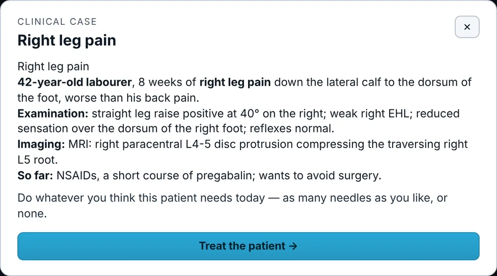
  • 33clinical cases with history, examination and imaging, from facet pain to cauda equina
  • 32multi-needle prescriptions, such as bilateral three-level RF neurotomy
  • 3grades of degeneration on top of normal anatomy, up to fusions, laminectomies, joint and disc replacements

Four ways to train

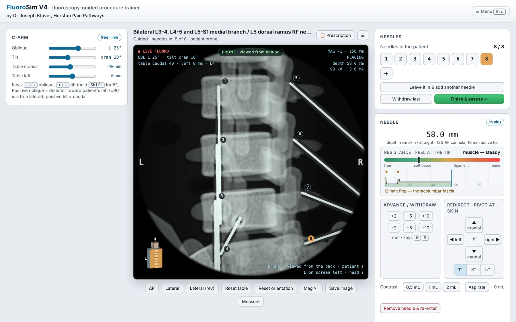
The procedure screen: C-arm controls on the left, the fluoroscopy image with eight numbered cannulae in the centre, and the needle controls on the right.

Work through a prescription

You're given a list of procedures — bilateral L3-4 to L5-S1 medial branch RF, say, or two-level transforaminal epidurals. Place every needle, leaving each in the patient while you place the next, then all of them are assessed together.

Go back and reposition any needle by its number at any point.

How a placement is assessed

  1. Get the view

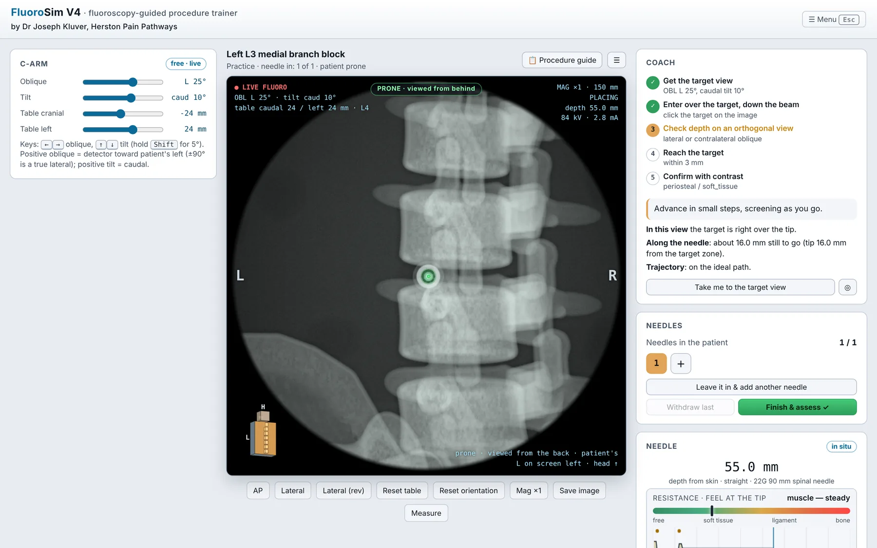
    Oblique and tilt the C-arm until the target is where the technique says it should be.

  2. Enter down the beam

    Start directly over the target, so the needle runs as a dot rather than a line.

  3. Check depth

    One view can't show depth: confirm on a lateral or contralateral oblique.

  4. Reach the target

    Feel the tissue planes change, touch bone where you should, and stop.

  5. Confirm with contrast

    The spread shows whether you're in the joint, the epidural space, a vessel or muscle.

  6. Get your feedback

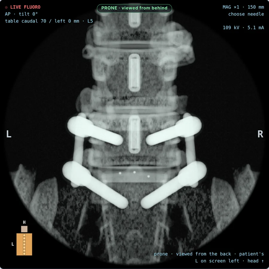
    Placement, risk and efficacy, with the ideal path drawn beside every needle.

Patients aren't textbook spines

Turn on degeneration and each patient gets their own: disc space narrowing, osteophytes, facet arthropathy, and at the severe end previous surgery — fusions, laminectomies, disc and joint replacements, cement augmentation and stimulator leads. The target always stays reachable; the path to it changes.

Eponymous conditions can be switched on too: Baastrup's disease, Bertolotti's syndrome and Maigne's syndrome, with the anatomy and the injections that treat them.

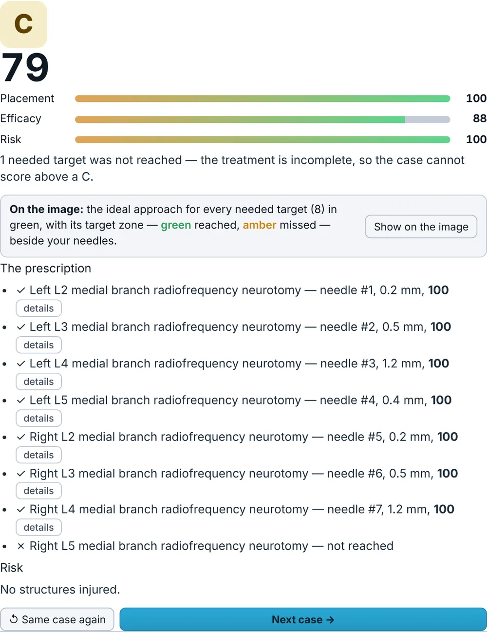
AP view of a normal lower lumbar spine.
NormalThe same patient, no degeneration.
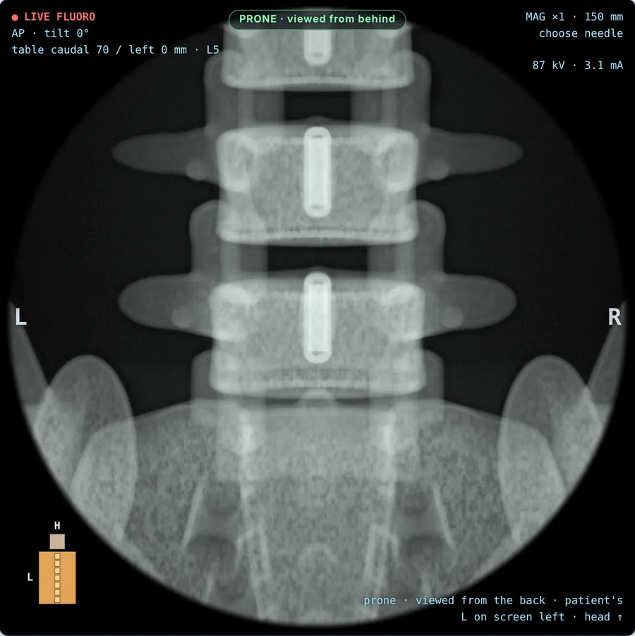
AP view of a severely degenerate lumbar spine with pedicle screws, rods and an interbody cage at L5-S1.
SevereL5-S1 instrumented fusion with an interbody cage, collapsed discs and osteophytes.

On a computer

Any current version of Chrome, Edge, Safari or Firefox. Keyboard shortcuts move the C-arm and screen, as a foot pedal would.

On an iPad

Touch controls for the C-arm and needle. In Safari, choose Share, then Add to Home Screen, and it opens full screen like an app.

Your progress stays with you

Settings and progress are saved in your browser on your own device. Nothing is uploaded and there's no account.

Made by a pain physician

FluoroSim is built by Dr Joseph Kluver, a specialist pain medicine physician (FANZCA, FFPMANZCA) at Herston Pain Pathways in Brisbane, for trainees and colleagues learning fluoroscopy-guided procedures.

Before you use it

FluoroSim is an educational tool. Its anatomy, tissue feel and radiographic appearances are simplified models, and its scores are training feedback, not an assessment of competence.

It is not a medical device. Don't use it to plan or guide the treatment of a real patient, and learn interventional procedures under qualified supervision.

The C-arm is warmed up.

Open the simulator